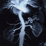
Magnetic Resonance Angiography
A Magnetic Resonance Angiography (MRA) is a type of Magnetic Resonance Imaging (MRI) scan that allows doctors to look at a patient’s blood vessels without using X-rays or other forms of radiation. MRAs allow the doctor to examine both the patient’s blow flow as well as the health of the blood vessels themselves. By using MRAs, doctors can discover any possible problems resulting from low blood flow and identify the source of the problem. MRAs can identify narrow regions of the patient’s blood vessels, damaged portions of the aorta in the heart, and blood clots in the brain that are caused by fat and calcium deposits. These are all serious conditions that require immediate medical attention. MRAs are therefore very important because they alert both the patient and the doctor to potential problems as soon as possible.
How Does A MagneticResonance Angiography Work?
When a Magnetic Resonance Angiography is performed, the patient is positioned so that either the patient’s entire body or a specific body part is inside the MRA machine. The machine itself is a large, cylindrical magnet that, when activated, produces a powerful magnetic field within the chamber. This magnetic field causes the nuclei of the hydrogen atoms within the human body to align themselves in much the same way that iron shavings align themselves when a magnet is waved over them. Radio waves are then used to scatter these hydrogen nuclei so that the magnetic field pulls them back into place. Metallic coils within the MRA’s chamber detect the movement of the nuclei, which the machine’s computer then converts into a digital image. In an MRA image, hydrogen-rich areas such as fat and muscle appear white while hydrogen-poor areas such as bone appear to be black. The doctor may also use a contrast dye to make blood vessels appear better in the image or to test blood flow. The computer can save MRA images and other information so that it can be reviewed or shared with the patient at a later time.
Health Effects
As MRAs do not use X-rays or other forms of radiation to view a patient’s blood vessels, there are virtually no health risks involved. However, MRAs can cause serious damage by effecting metallic objects both inside the MRA examination room as well as inside the patient. The strong magnetic field that the MRA machine produces is powerful enough to propel these items across the room or rip them out of the patient’s body. Health care workers must be very careful not to leave any metallic objects such as keys, pens, surgical equipment, or similar objects in the examination room. Wheelchairs, stretchers, oxygen bottles, and other clinical equipment in the room must be made of plastic or stainless steel. Likewise, the patient needs to make sure that his/her doctor is fully aware of any metallic instruments such as clips, screws, bolts, pins, or plates inside his/her body as well as electronic devices such as pacemakers. As patients can sometimes forget about specific objects within their body or never be made aware of them in the first place, a full surgical history should be verified before an MRA is performed.
Preparation
Many patients feel nervous before an MRA. This is completely normal and understood. MRAs require the user to lay very still inside of a very small, enclosed chamber and the thought of metal objects piercing their skin can scare any patient before their procedure. Fortunately, many modern MRAs are open in order to prevent any bouts of claustrophobia, and rigorous exam procedures leave little room for metal objects to injure the patient or the machine. As a result, the patient can simply concentrate on staying calm and relaxed throughout their procedure.


Comments - No Responses to “Magnetic Resonance Angiography”
Sorry but comments are closed at this time.